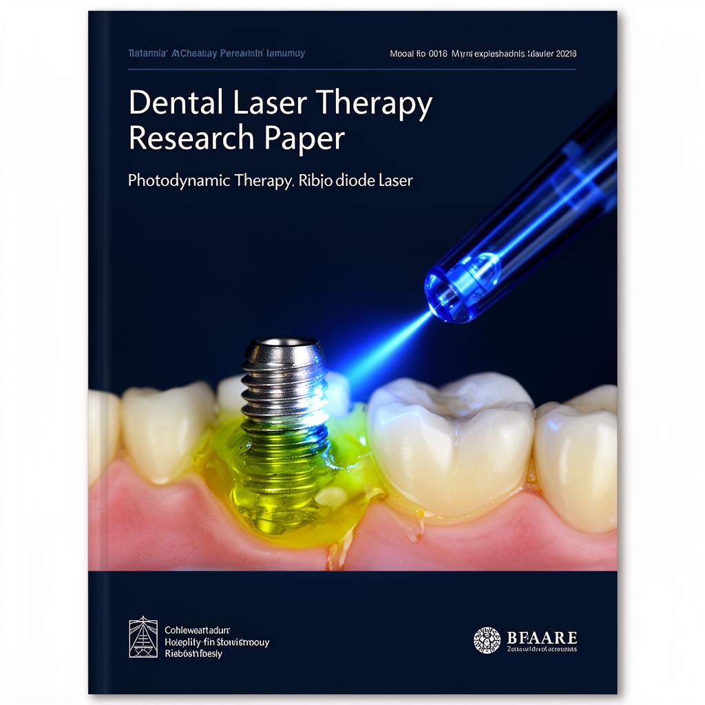
A Novel Technique for Disinfection Treatment of Contaminated Dental Implant Surface Using 0.1% Riboflavin and 445 nm Diode Laser — An In Vitro Study
Peri-implantitis remains one of the most challenging complications in modern implant dentistry. Despite decades of research and a wide range of available treatment protocols, no single method has been shown to completely eradicate the microbial biofilm that accumulates on implant surfaces and drives progressive bone loss.
A 2022 study published in Bioengineering (Morelato et al., co-authored by Prof. Muhamed Ajanović of the University of Sarajevo Faculty of Dentistry) investigated a novel approach: combining 0.1% riboflavin as a photosensitizing agent with a 445 nm blue diode laser, and comparing its antimicrobial efficacy against the more commonly used methylene blue and 660 nm red laser protocol. The results are clinically relevant and worth understanding in depth.
The Problem — Why Decontaminating Implant Surfaces Is So Difficult
Titanium implant surfaces are engineered for osseointegration — roughened, sandblasted, and acid-etched to maximize bone contact. The same surface topography that promotes osseointegration also makes decontamination exceptionally difficult. The micro- and macromorphology of implant threads creates areas that are essentially inaccessible to mechanical instruments.
More than 20 species of micro-organisms have been identified as peri-implant pathogens. Among the most significant are Staphylococcus aureus — an early colonizer with a high affinity for titanium surfaces — and Candida albicans, a fungal species capable of forming thick biofilms that are particularly resistant to standard chemical disinfection.
Why Chlorhexidine Alone Is Not Enough
Chlorhexidine (CHX) at 0.2% concentration is widely used for implant surface decontamination and remains the positive control against which most new approaches are benchmarked. It is effective — but not completely so. Anatomical limitations, the complexity of implant surface geometry, and the intrinsic resistance of mature biofilms mean that CHX cannot reliably eliminate all peri-implant pathogens.
Photodynamic therapy (PDT) has emerged as a promising adjunct — and the question being asked by researchers is no longer whether it works, but which protocol works best.
What Is Photodynamic Therapy in the Context of Implant Dentistry?
Antimicrobial PDT is based on a photochemical reaction involving three components: a photosensitizing agent, light at a specific wavelength, and oxygen. When a photosensitizer binds to microbial cells and is activated by light of the appropriate wavelength, it generates reactive oxygen species (ROS) that disrupt bacterial cell walls and normal cellular metabolism, leading to cell damage or death.
The mechanism is clinically important for several reasons:
PDT does not damage host cells, which have enzymatic defenses against oxidative stress (catalase, superoxide dismutase)
Microbial resistance to PDT is unlikely to develop, since the bactericidal effect is mediated through oxygen radical action on cellular components rather than through a specific biochemical pathway
Photosensitizing agents can penetrate pores and surface irregularities on implant surfaces that are inaccessible to mechanical instruments
The Standard Protocol — Methylene Blue and 660 nm Red Light
The most widely used PDT protocol in clinical dentistry combines methylene blue (MB) as the photosensitizer with red light in the 600–660 nm range. An American Academy of Periodontology systematic review concluded that PDT with methylene or toluidine blue can provide clinical improvements comparable to conventional periodontal therapy for both periodontitis and peri-implantitis patients.
However, methylene blue has a significant cosmetic drawback: it causes visible blue discoloration of the surrounding soft tissues, which limits its practical use in the aesthetic zone — precisely where peri-implantitis around anterior implants requires treatment.
The Novel Protocol — Riboflavin and 445 nm Blue Light
Riboflavin (vitamin B2) is a naturally occurring yellow pigment present in food. When used as a photosensitizer and activated by blue light at approximately 445 nm, it generates reactive oxygen species through both type I (electron transfer) and type II (energy transfer) photochemical reactions.
Its key clinical advantage is cosmetic: riboflavin does not cause the soft tissue discoloration associated with methylene or toluidine blue, making it particularly suitable for anterior aesthetic cases.
The protocol had previously been tested in endodontics (Katalinić et al., 2019) with promising results. The 2022 Morelato study was the first to apply it specifically to dental implant surfaces contaminated with a bacterial-fungal biofilm.
Study Design — How It Was Tested
The study used 80 titanium dental implants (GC Aadva Standard Implants, 4.0 mm × 10 mm) contaminated with a mixed Staphylococcus aureus and Candida albicans biofilm over 14 days under aerobic conditions. Biofilm formation was confirmed using scanning electron microscopy (SEM).
The implants were randomly divided into four groups of 20:
The Four Groups
Negative Control (NC) — No treatment. Used to establish baseline microbial load.
Positive Control (PC) — 0.2% chlorhexidine solution applied with a sterile cotton pellet using brushing movements along the implant threads for 60 seconds.
PDT1 (660 nm + Methylene Blue) — 0.1% methylene blue applied for 60 seconds, rinsed with saline, then irradiated with a 660 nm diode laser (SiroLaser Blue, Dentsply Sirona) at 124.34 W/cm², energy density 1240 J/cm², continuous wave mode, 60 seconds.
PDT2 (445 nm + Riboflavin) — 0.1% riboflavin applied for 60 seconds, rinsed with saline, then irradiated with a 445 nm diode laser at 124.34 W/cm², energy density 1.24 J/cm², pulsed mode at 100 Hz, 60 seconds.
Colony forming units (CFUs) for both S. aureus and C. albicans were counted after 48 hours of incubation, and SEM analysis was performed on one randomly selected implant from each group.
Results — What the Data Showed
Staphylococcus aureus Reduction
The negative control group showed a median CFU count of 3.2 × 10⁶ — representing the full bacterial load of untreated biofilm. Both PDT groups showed dramatic reductions that were statistically significant compared to the NC group (p < 0.001). Critically, there was no statistically significant difference between either PDT group and the CHX positive control group, and no statistically significant difference between PDT1 and PDT2 (p = 0.55).
Candida albicans Reduction
Results for C. albicans followed the same pattern. The NC group showed a median of 1.5 × 10³ CFU. Both PDT protocols produced reductions comparable to 0.2% CHX (p = 0.15 for PDT1 vs PC; p = 0.38 for PDT2 vs PC), and no significant difference was observed between PDT1 and PDT2 (p = 0.49).
SEM Findings
Scanning electron microscopy confirmed dense, evenly distributed biofilm on untreated implants. In the PC, PDT1, and PDT2 groups, only scattered single micro-organisms or small groups could be found on the implant surface — consistent with significant but not complete elimination. This finding aligns with a systematic review by Alasqah et al., which concluded that PDT reduces bacterial viability but cannot completely disrupt implant surface biofilm.
Clinical Implications — What This Means for Practice
Riboflavin PDT Is a Viable Alternative in the Aesthetic Zone
The most immediately actionable finding is that the riboflavin/445 nm protocol achieved antimicrobial efficacy equivalent to the established methylene blue/660 nm protocol, without the tissue discoloration that makes methylene blue unsuitable for anterior aesthetic cases. For clinicians treating peri-implantitis around visible anterior implants, this represents a meaningful clinical option.
PDT Should Be Used as an Adjunct, Not a Replacement
Both PDT protocols — and chlorhexidine — left some micro-organisms detectable on implant surfaces by SEM. The consistent conclusion across the literature is that PDT cannot replace mechanical debridement; it should be used in combination with it. The value of PDT lies in reaching areas of the implant surface that instruments cannot, reducing the residual bacterial load after mechanical cleaning.
Safety — Thermal Considerations
The use of laser energy on titanium implant surfaces carries a theoretical risk of thermal damage to adjacent tissues. For the 445 nm protocol used in this study (320 µm fiber, 100 mW, pulsed mode 100 Hz, 60 seconds), thermal risk was considered low. Referenced research by Deppe et al. demonstrated that a 445 nm laser at output powers of 100 mW in pulsed mode produces temperature rises within safe limits. Constant movement of the fiber tip during treatment further reduces thermal risk.
Resistance and Safety Profile
Neither photodynamic protocol poses any known risk of inducing antimicrobial resistance — an increasingly important consideration as antibiotic and antiseptic resistance becomes a clinical and public health concern.
Limitations and Future Directions
The authors appropriately acknowledge several limitations. This was an in vitro study, and the conditions of the oral cavity — salivary flow, immune response, anaerobic microenvironments, and limited accessibility — cannot be replicated in a laboratory setting. The concentrations and incubation times used may require optimization for clinical application.
Future research should focus on:
In vivo and randomized controlled clinical studies comparing riboflavin/445 nm PDT against standard protocols in peri-implantitis patients
Testing different riboflavin concentrations and formulations — the authors note that riboflavin-5'-phosphate (E101a), which has better solubility, may offer practical advantages over standard riboflavin
Evaluating the effect of repeated PDT applications, since a single 60-second treatment achieved significant but not complete bacterial elimination
Key Takeaways for Clinicians
The 2022 Morelato study provides in vitro evidence that a novel PDT protocol using 0.1% riboflavin and a 445 nm blue diode laser is as effective as the established methylene blue/660 nm protocol for reducing S. aureus and C. albicans biofilm on titanium implant surfaces, and comparable to 0.2% chlorhexidine.
For daily clinical practice, the key points are:
PDT is a validated adjunct to mechanical debridement in peri-implantitis treatment — not a replacement for it
Riboflavin/blue light offers the same antimicrobial efficacy as methylene blue/red light, with the advantage of no aesthetic soft tissue staining
A single 60-second treatment produces significant microbial reduction; repeated treatment may be more effective
The safety profile of the 445 nm laser at moderate parameters is well-supported in the literature
As peri-implantitis prevalence continues to rise alongside the global increase in implant placement, validated, practical decontamination protocols matter. This research adds to the evidence base for blue-light PDT as a clinically relevant option — particularly in aesthetic cases where the standard protocol's discoloration side effects are unacceptable.
This article is based on: Morelato L, Budimir A, Smojver I, Katalinić I, Vuletić M, Ajanović M, Gabrić D. A Novel Technique for Disinfection Treatment of Contaminated Dental Implant Surface Using 0.1% Riboflavin and 445 nm Diode Laser — An In Vitro Study. Bioengineering. 2022;9(7):308. https://doi.org/10.3390/bioengineering9070308
Interested in advancing your implant knowledge? Browse our clinical programs at european-academy.com/programs.